
Skull Development: How the Human Skull Grows from Birth to Adulthood
The human skull changes more than almost any other part of the skeleton over a lifetime. A baby’s skull is not the solid, fused structure of an adult; it is made of separate bony plates joined by soft, flexible tissue, with gaps called fontanelles that allow the skull to flex during birth and grow rapidly in infancy. This guide explains how the skull develops, from the newborn’s soft spots through the gradual fusion of the bones to the fully formed adult skull. It is a clear, factual guide to the anatomy of skull growth.
This is a companion to our complete guide to the human skull, focused on how the skull develops and grows.
A note before we begin: this article explains normal skull development as anatomy and education. It is not medical advice. If you have any concern about a child’s head shape or development, speak to a doctor or health visitor, who can examine the individual child and give proper guidance.
Why the baby skull is different
A newborn’s skull is strikingly different from an adult’s, and for good reason. It is built to do two things an adult skull does not: pass safely through the birth canal and accommodate the astonishingly fast growth of the brain in the first years of life. To do both, it cannot be a single rigid case.
Instead, a baby’s skull is made of separate bony plates that are not yet joined together. Between them lie bands of tough, flexible tissue, and at certain points these widen into soft gaps called fontanelles. This construction lets the plates move slightly so the head can change shape to fit through the birth canal, and it lets the skull expand as the brain grows. The adult skull’s rigid, protective strength is traded, in infancy, for flexibility and room to grow.
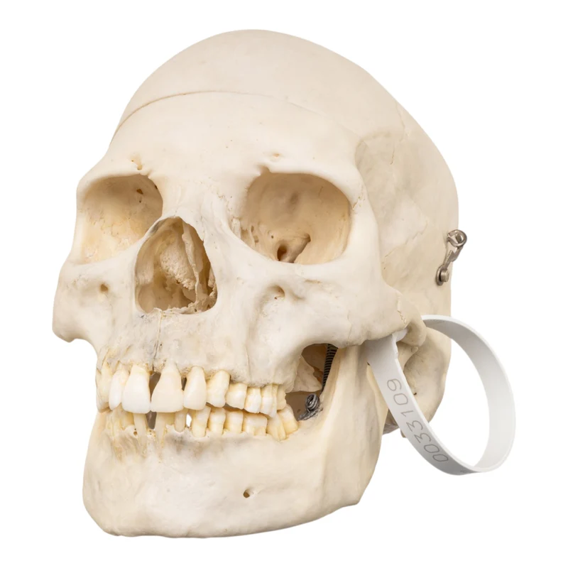
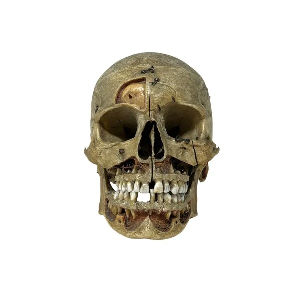
How skull bone forms: the two pathways
The skull is unusual in that its bones form in two different ways, and understanding this explains a lot about its development. Anatomists distinguish two processes of bone formation in the skull.
The flat bones of the cranial vault, the frontal, parietal, and parts of the occipital and temporal bones, form through a process called intramembranous ossification. Here, bone develops directly within a sheet of membrane, spreading outward from centers of ossification. Because these plates grow outward from their centers and only meet later, gaps naturally remain between them in early life, and those gaps are the fontanelles. This is precisely why the newborn skull has soft spots: the vault bones are still spreading toward one another.
The bones of the cranial base form through a different process, called endochondral ossification, in which bone replaces an earlier cartilage model. This is the same process that forms most of the long bones of the body. The base of the skull therefore develops on a slightly different pattern from the vault above it. Together, these two pathways build the complete skull, and the difference between them is a core concept in the study of skull development.
What are fontanelles? The soft spots explained
Fontanelles are the soft spots on a baby’s head, the gaps between the developing skull bones covered by tough membrane rather than bone. They are a completely normal and essential part of skull development, and there are several of them.
The two best known are
- The anterior fontanelle. The larger, diamond-shaped soft spot toward the front and top of the head, where the frontal and parietal bones will eventually meet. This is the “soft spot” most people know.
- The posterior fontanelle. A smaller, triangular soft spot toward the back of the head, where the parietal bones meet the occipital bone.
There are also smaller fontanelles on the sides of the skull. Though they feel delicate, fontanelles are covered by a tough, protective membrane and are more robust than they appear. They serve two purposes: they let the skull bones overlap slightly during birth, and they give the brain room to grow rapidly afterwards. As the skull develops, the fontanelles gradually close as bone fills in the gaps, a normal process covered below.
When do the fontanelles close?
The fontanelles close on a general timeline as the skull bones grow toward each other, though the exact timing varies from child to child. As a broad guide, the posterior fontanelle closes first, usually within the first few months, while the larger anterior fontanelle closes later, often somewhere in the first couple of years.
It is important to understand that this timing is a general pattern, not a fixed rule, and there is natural variation between healthy children. The closing of the fontanelles is one of several markers of normal development that health professionals monitor at routine check-ups. If you ever have a question about whether a child’s fontanelles are closing as expected, that is exactly the kind of thing to raise with a doctor or health visitor, who can assess the individual child. This article describes the general anatomy, not any particular case.
How the skull bones fuse: suture development
As covered in our skull anatomy guide, the adult skull bones are joined by immovable joints called sutures. In development, these sutures begin as the flexible tissue between the separate infant bones, and they change over many years.
In a newborn, the sutures are wide and mobile, allowing the plates to shift. Through infancy and childhood, the bones grow at their edges, the fontanelles fill in, and the sutures narrow into the interlocking seams of the mature skull. But the sutures do not fully fuse for a long time. They remain as visible joints throughout childhood and adulthood, and the bone across them continues to knit gradually over decades. This very slow, lifelong fusion is why sutures remain identifiable on an adult skull and why, as we will see, forensic scientists can use their degree of fusion to help estimate age.
One suture worth noting is the metopic suture, which runs down the midline of the frontal bone. In most people it fuses in early childhood, so the frontal bone becomes a single bone. In some people it persists into adulthood as a normal variant, still visible as a line down the forehead of the skull.
How the skull grows in size and shape
Beyond the closing of gaps, the whole skull grows dramatically in size and changes in proportion as a person develops. The growth is driven largely by the brain: as the brain expands, the cranial bones grow at their edges to keep pace, which is why the fontanelles and sutures are so important in early life.
The proportions change too. A baby’s skull has a very large cranium relative to a small face, which is part of why infant features look the way they do. As a child grows, the face, including the jaws and the air-filled sinuses, develops and enlarges, so the balance between the braincase and the face shifts toward the adult proportions. The teeth also develop in stages, from the milk teeth to the adult set, reshaping the jaws as they come through. By adulthood, the skull has reached its full size and its mature proportions, with a smaller cranium-to-face ratio than in infancy.
Baby head shape and why it varies
A newborn’s head shape often looks uneven at first, and understanding why is part of understanding skull development. Because the infant skull bones are separate and mobile, the head can be temporarily molded during birth, sometimes emerging with a slightly elongated or asymmetric shape. This is common and usually settles in the early weeks as the skull rounds out.
In the early months, because the bones are still soft and mobile, a baby’s head shape can also be influenced by how they lie and rest. This is normal anatomy at work: the same flexibility that allows birth and brain growth also makes the young skull responsive to pressure. Head shape is another thing health professionals routinely monitor. Because every child is different, and because head shape can occasionally reflect something that needs attention, any specific concern about a baby’s head shape should always go to a doctor or health visitor rather than a general article. What this guide offers is the underlying anatomy: a young skull is designed to be flexible, and that flexibility is why head shape can vary in the early months.
Skull development conditions: a note
Because skull development is so important, a number of medical conditions relate to how the skull grows, and you may come across their names. Terms like craniosynostosis (where sutures fuse too early) and positional head-shape differences are medical diagnoses, not topics this article can assess.
We mention them only so you recognize that these are medical matters. If you are researching a specific condition or worried about a particular child, the right source is a qualified medical professional, a pediatrician, GP, or specialist, who can examine the child and give accurate, personal guidance. No general article, including this one, can or should substitute for that. Our focus here is the normal anatomy of how the skull develops.
Teeth and the developing jaw
The development of the teeth is a major part of how the skull grows, and it reshapes the jaws over many years. A newborn typically has no visible teeth, but the jaws already hold the developing buds of both the milk teeth and, beneath them, the future adult teeth.
The milk teeth, also called primary or deciduous teeth, erupt through infancy and early childhood, a set of twenty. As the child grows, these are gradually lost and replaced by the larger adult set, up to thirty-two teeth, including the wisdom teeth, which are the last to appear, often in early adulthood. Each stage changes the shape and size of the jaws, since the bone must grow to house progressively larger teeth. This is why the lower face and jaws enlarge so much between infancy and adulthood, shifting the proportions of the whole skull. The predictable sequence in which teeth develop and erupt is, like suture fusion, one of the markers used to estimate age from a skull, which is why dental development is studied so closely in both anatomy and forensics.
Why skull development matters
Understanding how the skull develops is not just academic; it underpins several fields and explains things we all notice. In medicine, knowing the normal timeline of fontanelle closure and suture fusion is how professionals recognize healthy development, which is why these are checked at routine infant appointments. In forensic science, the sequence of development is what allows an expert to estimate age from remains, as we explore in our forensic skull analysis guide.
For anyone studying anatomy or biology, skull development is a perfect example of how form follows function: the newborn skull is flexible because it must be, and the adult skull is rigid because it must be, and the whole process of change carries the body from one need to the other. And for artists and anthropologists, understanding how the skull’s proportions shift with age explains why children and adults look so different, and why a skull can reveal so much about the person it belonged to. Whichever field draws you in, the development of the skull is one of the most elegant stories in human anatomy.
The adult skull: the end of development
By adulthood, the dramatic development of the skull is complete, and the result is the structure described in our human skull anatomy guide. The fontanelles have long since closed, the separate infant plates have grown together into the 22 bones of the mature skull, and the sutures have narrowed into their interlocking adult form.
But the skull is never entirely static. The sutures continue their very slow fusion across adult life, and bone continues to remodel gradually, as it does throughout the skeleton. In old age, further subtle changes occur, including changes to the jaw, particularly after the loss of teeth, which alters the shape of the mandible and the lower face. So while the major development is finished by adulthood, the skull continues to change quietly across the whole of life, from the flexible, soft-spotted skull of the newborn to the fully knit skull of old age.
Reading age from the skull
One of the most fascinating consequences of skull development is that a skull records the age of its owner. Because the stages of development happen on a broad timeline, the features of a skull reveal roughly how old a person was.
In a young skull, the unfused sutures, open or recently closed fontanelles, and developing teeth all point to a child. In an adult skull, the degree of suture fusion and the state of the teeth help narrow the age further. This is exactly the principle forensic anthropologists use to estimate age from skeletal remains, and it works precisely because skull development follows such a consistent, well-understood sequence. We explore this in our forensic skull analysis guide. It is a striking reminder that the skull is not a fixed object but a record of a life’s development.
Infant skull vs. adult skull: the key differences
Placing an infant skull and an adult skull side by side is the clearest way to see the whole story of development, and the differences are striking. This comparison is a staple of anatomy teaching for exactly that reason.
| Feature | Infant skull | Adult skull |
|---|---|---|
| Fontanelles | Present (open soft spots) | Closed, filled with bone |
| Sutures | Wide, mobile | Narrow, interlocked |
| Separate plates | Yes, not yet fused. | Grown into 22 bones |
| Cranium-to-face ratio | Large cranium, small face | Smaller cranium relative to face |
| Teeth | None erupted, or milk teeth. | Full adult set (unless lost) |
| Flexibility | Flexible, can be molded | Rigid and fixed |
| Sinuses | Small or undeveloped | Fully developed |
The infant skull is built for birth and growth: flexible, gap-filled, and dominated by the braincase. The adult skull is built for protection: rigid, fully knit, with a developed face and sinuses. Everything between the two, the closing fontanelles, the fusing sutures, and the growing face and erupting teeth, is the process of development this guide describes. Seeing both together turns that timeline into something you can grasp at a glance, which is why teaching collections value specimens of different ages.
Studying skull development
For students of anatomy, biology, and forensics, understanding skull development is essential, and it is far easier to grasp with real or model specimens in hand. A fetal or infant skull specimen or model shows the separate plates and the fontanelles vividly, in a way no diagram can. Comparing an infant skull with an adult skull side by side makes the whole story of development instantly clear.
This is why comparative and developmental specimens are valued in teaching collections. Seeing the wide-open fontanelles of a newborn skull, then the fully fused adult skull, turns an abstract timeline into something concrete. Explore our human skull collection and our detailed anatomy guide to study the structure in depth.
Frequently asked questions
Why does a baby have soft spots on its head?
The soft spots, called fontanelles, are gaps between the developing skull bones covered by a tough membrane. They let the skull flex during birth and give the brain room to grow rapidly in infancy. They are a normal, essential part of skull development.
How many bones are in a baby’s skull?
A baby’s skull has more separate pieces than an adult’s, because the bones that will later fuse are still separate plates. As the child grows, these grow together into the 22 bones of the adult skull.
When do the fontanelles close?
As a general guide, the smaller posterior fontanelle closes within the first few months and the larger anterior fontanelle closes later, often within the first couple of years. Timing varies between healthy children, and a doctor monitors this at routine check-ups.
Why is a newborn’s head sometimes an odd shape?
Because the infant skull bones are separate and mobile, the head can be temporarily molded during birth, sometimes looking elongated or uneven at first. This usually settles in the early weeks. Any specific concern should go to a doctor.
Do skull bones fuse completely?
The separate infant plates grow together into the 22 adult bones, but the sutures joining them remain visible and continue to fuse very slowly across adult life. This is why sutures can still be seen on an adult skull.
When is the skull fully developed?
The major development is essentially complete by adulthood, when the fontanelles have closed and the bones have grown together. The sutures continue a very slow fusion, and the skull keeps remodeling subtly throughout life.
What is the metopic suture?
The metopic suture runs down the midline of the frontal bone. It usually fuses in early childhood so the frontal bone becomes one, but in some people it persists into adulthood as a normal variant.
Should I worry about my baby’s head shape or soft spot?
This article explains normal anatomy only and cannot assess any individual child. If you have any concern about a baby’s head shape, soft spot, or development, speak to a doctor or health visitor, who can examine the child and give proper advice.
Understand the skull’s remarkable journey.
The development of the skull, from the flexible, soft-spotted skull of a newborn to the fully formed skull of an adult, is one of the most remarkable stories in human anatomy. It explains the soft spots, the changing head shape, the slow fusion of the sutures, and even how age can be read from a skull. To explore further, read our complete guide to the human skull and detailed anatomy guide, or browse our human skull collection. And remember: for any question about a real child’s development, a doctor is always the right source. Every specimen we sell is responsibly sourced and ships worldwide in 3 to 5 days.