Skull Anatomy: A Complete Guide to the Bones, Sutures, and Structure
Skull anatomy describes how the 22 bones of the human skull fit together to protect the brain and form the face. The skull divides into the cranium, made of 8 bones that enclose the brain, and the facial skeleton, made of 14 bones that shape the face and jaw. This guide is a detailed, labelled walkthrough of the whole structure: every bone and where it sits, the sutures that join them, the openings that carry nerves and vessels, and how the skull looks from each anatomical view. It is written for students, artists, and anyone who wants to understand the skull in depth.
This is the technical companion to our complete guide to the human skull, which gives the wider overview.
Skull anatomy at a glance
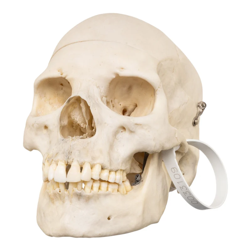
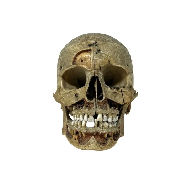
Before the detail, here is the structure in summary. The human skull has 22 bones, arranged in two groups.
| Region | Bones | Role |
|---|---|---|
| Cranium (neurocranium) | 8 | Encloses and protects the brain |
| Facial skeleton (viscerocranium) | 14 | Forms the face, orbits, nose, and jaw |
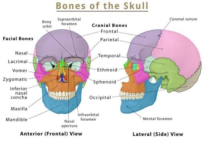
The 8 cranial bones are the frontal, occipital, sphenoid, and ethmoid (single bones) plus the paired parietal and temporal bones. The 14 facial bones are the paired maxillae, zygomatic, nasal, lacrimal, palatine, and inferior nasal conchae, plus the single vomer and mandible. The bones are joined by immovable joints called sutures, with one exception: the mandible, which moves at the jaw joint. This division into a protective braincase and a functional face is the single most important idea in skull anatomy, and everything below expands on this framework.
The cranial bones in detail
The cranium forms the protective vault around the brain. Here is each of its eight bones, in anatomical detail.
Frontal bone
The frontal bone forms the forehead, the roof of the orbits (eye sockets), and the front of the cranial floor. It contains the frontal sinuses. In infancy it develops as two halves joined by the metopic suture, which usually fuses in early childhood, though it can persist into adulthood as a normal variant.
Parietal bones
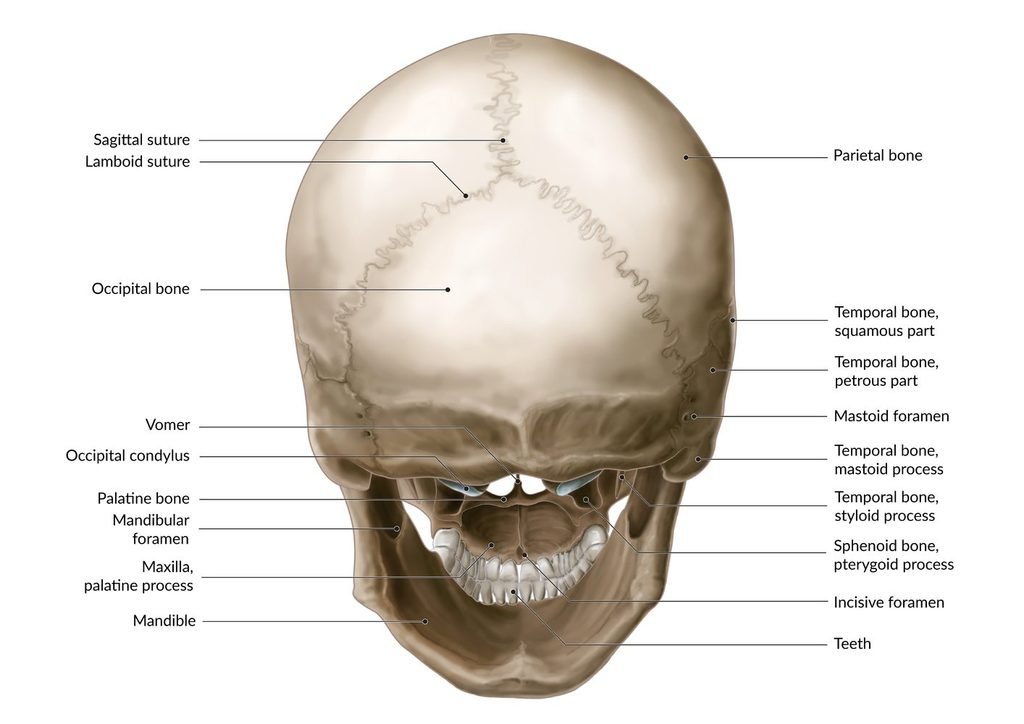
The two parietal bones form most of the roof and upper sides of the cranium. They meet each other at the midline along the sagittal suture, meet the frontal bone at the coronal suture, and meet the occipital bone at the lambdoid suture. They are the large, smooth plates that give the top of the head its dome.
Temporal bones
The two temporal bones sit at the lower sides of the skull. Each is complex, housing the middle and inner ear, and each carries the mandibular fossa, the socket that receives the jaw. The mastoid process, the bony bump behind the ear, is part of the temporal bone, as is the zygomatic process that helps form the cheek arch.
Occipital bone
The occipital bone forms the back and much of the base of the skull. Its defining feature is the foramen magnum, the large opening through which the brainstem passes to become the spinal cord. On either side of it are the occipital condyles, which articulate with the first vertebra of the neck and allow the nodding movement of the head.
Sphenoid bone
The sphenoid is a single, butterfly-shaped bone spanning the cranial floor. It articulates with almost every other cranial bone, earning it the name “keystone of the skull.” Its central saddle, the sella turcica, cradles the pituitary gland. The sphenoid contributes to the orbits, the nasal cavity, and the base of the braincase.
Ethmoid bone
The ethmoid is a small, light, spongy bone between the orbits, forming part of the nasal cavity roof and the medial orbital walls. Its cribriform plate is perforated by tiny holes through which the olfactory nerves of smell pass from the nose into the brain.
The facial bones in detail
The facial skeleton shapes the face, holds the teeth, and frames the eyes and nose. Here is each of its fourteen bones.
Maxillae
The two maxillae fuse at the midline to form the upper jaw. They hold the upper teeth and form the front of the hard palate, the floor of the nose, and part of the orbits. They are the largest immovable facial bones and central to the face’s structure.
Zygomatic bones
The two zygomatic bones are the cheekbones. Each forms the prominence of the cheek and part of the lateral wall and floor of the orbit and joins the temporal bone to complete the zygomatic arch.
Nasal bones
The two small nasal bones form the bridge of the nose, joining the frontal bone above and the maxillae to the sides. Below them, the shape of the nose is formed by cartilage, not bone.
Lacrimal bones
The two lacrimal bones are the smallest bones of the face, thin and fragile, sitting in the medial wall of each orbit. They carry a groove that forms part of the passage draining tears from the eye to the nose.
Palatine bones
The two L-shaped palatine bones form the back of the hard palate and part of the nasal cavity and orbit floor. They sit behind the maxillae, out of direct view.
Inferior nasal conchae
The two inferior nasal conchae are thin, scroll-like bones projecting into the nasal cavity. They increase its surface area, helping to warm and filter inhaled air.
Vomer
The vomer is a single, thin, flat bone forming the lower and back part of the nasal septum, the wall dividing the two nostrils.
Mandible
The mandible is the lower jaw and the only freely movable bone of the skull. It holds the lower teeth and meets the temporal bones at the temporomandibular joints. Its parts, the body, the angle, and the vertical rami rising to the joint, are important landmarks. The ramus of the mandible is the upright portion on each side that connects to the skull.
The sutures of the skull
The bones of the skull are joined by sutures, interlocking immovable joints. These are important landmarks in anatomy, forensics, and surgery. The main sutures are
- Coronal suture. Runs side to side across the top of the skull, joining the frontal bone to the two parietal bones.
- Sagittal suture. Runs front to back along the midline, joining the two parietal bones to each other.
- Lambdoid suture. Runs across the back, joining the parietal bones to the occipital bone. Its name comes from its resemblance to the Greek letter lambda.
- Squamous sutures. Run along each side, joining the parietal bones to the temporal bones.
- Metopic (frontal) suture. The midline suture of the frontal bone is present in infancy and usually fused by early childhood, though sometimes persisting.
Key meeting points of these sutures have their own names. The bregma is where the coronal and sagittal sutures meet; the lambda is where the sagittal and lambdoid sutures meet. These begin life as the fontanelles, the soft spots of a baby’s skull, covered in our skull development guide.
The foramina and openings of the skull
The skull is pierced by numerous openings called foramina, which allow nerves and blood vessels to pass through. Understanding these is a core part of skull anatomy, particularly for medical and forensic study.
The largest opening is the foramen magnum in the occipital bone, through which the spinal cord connects to the brain. Other important foramina include the optic canals, which carry the optic nerves to the eyes; the various openings of the cranial base that transmit the cranial nerves; and small foramina in the facial bones that carry sensory nerves to the face, such as the supraorbital, infraorbital, and mental foramina, which lie roughly in a vertical line and are landmarks used in anatomy and dentistry.
Together with the air-filled paranasal sinuses in the frontal, sphenoid, ethmoid, and maxillary bones, these openings make the skull far lighter than a solid structure of the same size, without sacrificing its strength.
The anatomical views of the skull
Anatomists describe the skull from standard viewpoints, and knowing these helps you read any labelled diagram. Each view reveals different features.
Anterior view (from the front). Shows the forehead, orbits, nasal opening, cheekbones, and upper and lower jaws. This is the view most people picture, and it displays the facial skeleton clearly.
Lateral view (from the side). Shows the profile: the cranial vault, the temporal region, the zygomatic arch, and the relationship of the jaw to the rest of the skull. The sutures are clearly visible from the side.
Posterior view (from behind). Shows the back of the parietal and occipital bones and the lambdoid suture.
Superior view (from above). Shows the top of the cranial vault and the coronal, sagittal, and lambdoid sutures meeting like a map.
Inferior view (from below, with the mandible removed). Shows the base of the skull: the hard palate, the foramen magnum, the occipital condyles, and the many foramina of the cranial base. This is the most complex view and the most useful for studying the openings.
Studying all five views together gives you a full three-dimensional understanding of the skull, which is exactly why anatomy courses and models present them side by side.
The regions of the cranial base
The floor of the cranium, the cranial base, is worth understanding on its own because it is one of the most intricate parts of skull anatomy. Viewed from inside, it forms three stepped levels called cranial fossae.
The anterior cranial fossa is the highest and most forward, supporting the frontal lobes of the brain. The middle cranial fossa sits lower, shaped by the sphenoid and temporal bones, and cradles the temporal lobes; it contains the sella turcica for the pituitary gland. The posterior cranial fossa is the deepest and most rearward, formed largely by the occipital bone, and holds the cerebellum and brainstem, with the foramen magnum at its center.
These three fossae, descending like shallow steps from front to back, are a key concept in neuroanatomy and surgery and a frequent subject of study for anyone learning the skull in depth.
Muscles and the skull
The skull is not just a passive case; it is covered in attachment points for muscles, and understanding these adds another layer to skull anatomy. Two groups matter most.
The muscles of mastication, the chewing muscles, attach the mandible to the skull and power the bite. The powerful temporalis muscle fans across the temporal region and lifts the jaw, while the masseter runs from the cheekbone to the angle of the mandible. Together they generate the considerable force of the human bite. You can feel both working if you clench your teeth and touch your temple and jaw.
The muscles of facial expression are unusual because many attach not to other bones but to the skin of the face. Anchored to the facial skeleton, they move the skin to create the enormous range of human expressions, from a smile to a frown. This is why the underlying shape of the facial bones so strongly determines how a face looks and moves, and why artists study the skull to understand the face built upon it. The facial muscles are a frequent subject of anatomical study in their own right.
The temporomandibular joint
The one movable joint of the skull deserves its own attention, because it is among the most used joints in the body. The temporomandibular joint, or TMJ, is where the mandible meets the temporal bone, just in front of each ear. It is a hinge-and-slide joint: it opens and closes like a hinge but also glides forward, which is what lets the jaw move up, down, side to side, and forward.
You use this joint constantly for chewing, speaking, yawning, and swallowing. A small disc of cartilage cushions the joint and lets it move smoothly. Because it is so heavily used and so complex, the TMJ is also a common site of dysfunction, which is why it is well known in dentistry and medicine. In skull anatomy, it is the single point where the otherwise rigid skull comes to life and moves.
How to learn and remember the skull bones
Learning the 22 bones of the skull is a rite of passage for anatomy students, and a few approaches make it easier. Grouping is the first key: learn the 8 cranial bones and the 14 facial bones as two separate sets rather than one long list. Within the facial bones, remember that six are paired and two are single (the vomer and mandible), which accounts for all fourteen.
Mnemonics help many learners. A common one for the cranial bones, “Old People From Texas Eat Spiders,” cues occipital, parietal, frontal, temporal, ethmoid, and sphenoid. Working with a physical skull or model is the most effective method of all, because handling the bones and tracing the sutures builds a three-dimensional memory that flat diagrams cannot. Testing yourself with a skull bones quiz, naming each bone and suture on an unlabelled diagram, quickly reveals what you know and what needs review. Repetition across all five anatomical views cements it.
Bones associated with the skull
A few bones sit close to the skull and are often discussed alongside it, even though they are not counted among the 22. Knowing them rounds out your understanding of the region.
The hyoid bone is a small, U-shaped bone in the neck, below the mandible. It is unusual because it does not directly join any other bone; instead, it is suspended by muscles and ligaments. It anchors the tongue and muscles of the throat and plays a role in swallowing and speech. Because it is not attached to the skull, it is counted separately.
The auditory ossicles, three tiny bones in each middle ear, the malleus, incus, and stapes, are the smallest bones in the entire body. They sit within the temporal bones and transmit sound vibrations to the inner ear. Although housed inside the skull, they are counted as their own group rather than as skull bones, which is why the standard skull total is 22 rather than 28.
Finally, the first vertebra of the neck, the atlas, meets the occipital condyles at the base of the skull to form the joint that lets you nod. While part of the spine rather than the skull, this junction is where the skull connects to the rest of the skeleton, completing the anatomical picture.
Studying skull anatomy
Because skull anatomy is complex and three-dimensional, it is difficult to learn from flat diagrams alone. This is why students, artists, and educators value a physical skull, whether a genuine specimen or an accurate replica, that can be turned, opened, and examined from every view.
A good anatomical skull model shows the sutures, the foramina, and the relationships between bones far more clearly than any illustration. Many models come apart to reveal the cranial base and interior or are color-coded by bone. For serious study of natural variation, a real specimen is unmatched. We cover the options in our guide to replica and model skulls, and you can browse genuine and replica specimens in our human skull collection.
Frequently asked questions
How many bones are in the skull?
The skull has 22 bones: 8 cranial bones that protect the brain and 14 facial bones that form the face and jaw. This excludes the six middle-ear ossicles and the hyoid bone, which are counted separately.
What are the main sutures of the skull?
The main sutures are the coronal (front to parietal), the sagittal (between the two parietal bones), the lambdoid (parietal to occipital), and the squamous (parietal to temporal). The metopic suture runs down the midline of the frontal bone in infancy.
What is the coronal suture?
The coronal suture is the joint that runs across the top of the skull from side to side, joining the frontal bone at the front to the two parietal bones behind it.
What is the foramen magnum?
The foramen magnum is the large opening in the base of the occipital bone through which the brainstem passes to become the spinal cord. It is the largest of the skull’s many foramina.
What is the ramus of the mandible?
The ramus is the upright portion of the lower jaw on each side, rising from the body of the mandible to the joint with the skull. There is a left and a right ramus.
What is the difference between cranial and facial bones?
Cranial bones (8) form the protective case around the brain. Facial bones (14) form the face, orbits, nasal cavity, and jaw. Together they make up the 22 bones of the skull.
Which is the only movable bone in the skull?
The mandible, or lower jaw, is the only freely movable bone of the skull. All the other bones are joined by immovable sutures.
What is the cranial base?
The cranial base is the floor of the cranium. Seen from inside, it forms three stepped levels, the anterior, middle, and posterior cranial fossae, that support different parts of the brain.
How do you learn the bones of the skull?
Learn the 8 cranial and 14 facial bones as two separate groups, use a mnemonic for the cranial bones, and study with a physical skull or model. Testing yourself on an unlabelled diagram, a skull bones quiz, quickly shows what you have mastered.
Master the anatomy of the skull.
Skull anatomy rewards close study: 22 bones, dozens of features, and five distinct views, all working together to protect the brain and shape the face. The best way to learn it is with a skull in hand, turning it to see how the pieces fit. Explore our complete guide to the human skull, our guide to replica and model skulls, or browse genuine and replica specimens in our human skull collection. Every specimen is responsibly sourced and ships worldwide in 3 to 5 days.